In all living cells, proteins are synthesized by ribosomes. . The ribosome is a large macromolecule with a folding asymmetric quarter structure, inspired by ribonucleic acids (ribosomal RNA) and proteins. In order to synthesize proteins, the ribosome must be secured:
1. A program that sets the order of picking up amino acid residues in the polypeptide lance of the protein.
2. Amino acid material, from which the protein will be used.
3. Energy.
The ribosome itself has a catalytic (enzymatic) function, responsible for the incorporation of peptide bonds and, apparently, polymerization of amino acid residues in the polypeptide lance of the protein.
The program, which sets the order of amino acid residues in the polypeptide lance of the protein, appears in the form of deoxyribonucleic acid (DNA), i.e., from the genome of the clitin. The synthesis of RNA lances is complementary to one of the DNA lances i, in such a way, exactly confirms the deoxyribonucleotide sequence of the other DNA lance in its ribonucleotide sequence. The process of such copying of a gene, which is modified by the enzyme RNA polymerase, removing the name of the transcription. RNA during the synthesis of the latter, especially in eukaryotic clitins, can be subject to a number of additional changes, called processing, in the course of which, they can be changed into small parts of the nucleotide sequence. RNA, which enters, enters far into the ribosomes as a program, which determines the amino acid sequence in the synthesized protein. It is called informational or "messenger" RNA (mRNA). In this way, the very transcription of genes and the adoption of mRNA ensure the flow of information from DNA to ribosomes.
The cob material, from which the protein will be, is amino acids. However, amino acids are not vicarious by the ribosome. In order to serve as a substrate for the ribosome, the amino acid must be activated for the participation of the resulting splitting of ATP and is accepted (covalently attached) by a special RNA molecule, called transfer or transfer RNA (tRNA), with additional help. Otrimani aminoacyl-tRNA is present in the ribosome as a substrate for protein synthesis. In addition, the energy of the chemical bond between the amino acid excess and tRNA is vicorated for the reaction of the peptide bond in the ribosome. Thus, the activation of amino acids and the adoption of aminoacyl-tRNA provide both material and energy for ribosomal protein synthesis.
The three streams (information, material and energy) are clustered in ribosomes. By accepting it, the ribosome transfers, or translates, genetic information from the nucleotide sequence of the mRNA to the sequence of the amino acid sequence of the synthesized polypeptide lancet protein. Yakshcho Vyavita Ts in molecular termns, then the ribosoma of the subganum of Lantsyug MRNA (rush to the sinking of non-schools), I have been tied to the soressine of the aminoacil-tribe. split mRNA. Also, the problem of the genetic code is to be blamed: which combinations of nucleotides determine, i.e., which code the skin with 20 amino acids, from which protein molecules will be formed?
The rotation of the ribosome of the lancet mRNA (or, in other words, the passage of the lancet of the mRNA through the ribosome) sets a strict timchasial order of entry into the ribosome of various aminoacyl-tRNAs, depending on the order of the expansion of the nucleotide combinations that encode, reduce the mRNA. The aminoacyl excess of the reversed aminoacyl-tRNA is covalently adjoined by the ribosome to the growing polypeptide lance. Deacylated tRNA is released from ribosomes at the sites. So sequentially, step by step, there will be a polypeptide lance of the protein (div. scheme 1).
Biosynthesis of protein.
Plastic exchange (assimilation and anabolism) - the sequence of reactions of biological synthesis. The name of this kind of exchange is the day of day: from speeches, like a clitin’s posture, speeches are settled, similar to the speeches of a clitin.
Let's look at one of the most important forms of plastic exchange - the biosynthesis of proteins. Biosynthesis of proteins found in all clitin pro-eukaryotes. Information about the primary structure (order of amino acids) of a protein molecule is encoded by the sequence of nucleotides in the DNA molecule division - gene.
The gene of a DNA molecule, which determines the order of amino acids in a protein molecule. Also, in the form of nucleotides in the gene, the order of amino acids in the polypeptide should be determined, that is. This is the primary structure, in view of which all other structures, the power and function of the protein molecule, lie in its own line.
The system for recording genetic information in DNA (i RNA) in the apparent sequence of nucleotides is called the genetic code. Tobto. genetic code unit (codon) - a triplet of nucleotides in DNA or RNA that encodes one amino acid.
Our genetic code includes 64 codons, of which 61 are coding and 3 are non-coding (terminator codons, which indicate the completion of the translation process).
Codon-terminators i - RNA: UAA, UAG, UGA, in DNA: ATT, ATC, ACT.
The translational process is initiated by the codon-initiator (AUG, DNA - TAC), which encodes the amino acid methionine. The cei codon is the first to enter the ribosome. By the way, methionine, although it is not transferring, as the first amino acid of this protein, is used.
The genetic code can be characteristic of power.
1. Universality - the code of the same for all organisms. One and the same triplet (codon) in any organism encodes the same amino acid.
2. Specificity - the skin codon codes for only one amino acid.
3. Virogenity - more amino acids can be encoded by the number of codons. There are 2 amino acids - methionine and tryptophan, which can only be used for one variant of the codon.
4. Mіzh genes є “different signs” - three special triplets (UAA, UAG, UGA), the skins of which indicate the synthesis of the polypeptide lanciug.
5. There are no middles of the gene of “different signs”.
In order to synthesize the protein, information about the sequence of nucleotides in its primary structure can be delivered to the ribosomes. This process includes two stages - transcription and translation.
Transcription(rewriting) information is taken by way of synthesis on one of the lances of the DNA molecule of the single-lancet RNA molecule, the sequence of nucleotides is exactly the same as the sequence of the nucleotides in the matrix - the polynucleotide lance of DNA.
Vaughn (i - RNA) is an intermediary that transmits information from DNA to the site of the selection of protein molecules in ribosomes. Synthesis of i RNA (transcription) is considered to be an offensive rank. Enzyme (RNA - polymerase) splits the sublingual lancet of DNA, and one of the lancets (coding) follows the principle of complementarity, RNA nucleotides are vibrated. The RNA molecule is synthesized in this way (matrix synthesis) and enters the cytoplasm, and small subunits of ribosomes are strung on one end.
Another step in protein biosynthesis is broadcast- tse translation of the sequence of nucleotides in the molecule and - RNA in the sequence of amino acids in the polypeptide. In prokaryotes, which cannot form a well-formed nucleus, ribosomes can bind to a newly created i-RNA molecule immediately after її dividing the DNA, or build up to the complete completion of її synthesis. In eukaryotes, i-RNA can be transported to the cobweb through the nuclear membrane into the cytoplasm. The transfer is caused by special proteins, which form a complex with an RNA molecule. The cream functions of transference of cytoplasmic proteins are protected by i - RNA from diy, which allows cytoplasmic enzymes.
In the cytoplasm, one of the ends of i - RNA (and on the one that initiates the synthesis of a molecule in the nucleus) enters the ribosome and initiates the synthesis of the polypeptide. In the world, passing through the RNA molecule, the ribosome translates triplet after triplet, successively adding amino acids to the growing end of the polypeptide lancet. The exact correspondence of the amino acid to the triplet code and RNA is ensured by t RNA.
Transport RNA (t-RNA) "bring" amino acids to the large subunit of the ribosome. The t-RNA molecule can be folded configuration. On some sites between complementary nucleotides, water bonds are established, and the molecule is shaped like a stable leaf. At the top of the folds, there is a triplet of free nucleotides (anticodon), which contains a single amino acid, and the base contains a chain of amino acids (Fig. 1).
Mal. 1. Scheme of transport RNA: 1 - water links; 2 - anticodon; 3-mіsce attached amino acids.
Skin t-RNA can only transfer its own amino acid. T-RNA is activated by special enzymes, adding its own amino acid and transporting it to the ribosome. In the middle of the ribosomes at the skin moment there are only two codons of i-RNA. Since the t-RNA anticodon is complementary to the i-RNA codon, there is a timely advent of t-RNA with an amino acid to i-RNA. Before the next codon comes another t-RNA, as if carrying its amino acid. Amino acids are mixed with the help of the large subunit of the ribosome, and with the help of enzymes, peptide bonds are established along with them. The linkage between the first amino acid and the її t-RNA, and the t-RNA goes from the ribosome behind the attacking amino acid. The ribosome moves one triplet, and the process is repeated. In this way, the molecule of the polypeptide is incrementally built up, in which amino acids are mixed in a sequence of sequences in the order of triplets that encode them (matrix synthesis) (Fig. 2).

Mal. 2. Scheme for protein synthesis: 1 – i-RNA; 2 - subunits of the ribosome; 3 – t-RNA with amino acids; 4 – t-RNA without amino acids; 5 - polypeptide; 6 - ta-RNA codon; 7-anticodon of tRNA.
One ribosome is capable of synthesizing a new polypeptide lance. Prote, often one molecule of i-RNA collapses a sprat of ribosomes. Such complexes are called polyribosomes. After the completion of the synthesis, the polypeptide lances are fused in the form of a matrix - i-RNA molecules, curl into a helix and gain a powerful (secondary, tertiary or quarter) structure. Ribosomes work even more efficiently: by stretching 1c, the bacterial ribosome converts the polypeptide lancet from 20 amino acids.
The biosynthesis of proteins (polypeptides) is a supra-adventitious folding and marvelous process. Biosynthesis of proteins actively occurs in all organs and tissues, including erythrocytes. A lot of clitin synthesize proteins for "export" (liver clitin, subscapular follicles), and in this way the stench will avenge even a large number of ribosomes. In animal cells, the number of ribosomes reaches 10 5, the diameter of the ribosome reaches 20 nm.
The process of protein synthesis takes place in the middle of the cells on the surface of the ribosomes, as complexes of two subunits with a sedimentation constant of 60S and 40S, which function as a single unit. In ribosomes, proteins become 30-35% and ribosomal RNA - 65-70%. Ribosomes have aminoacyl and peptidyl clefts. The first one serves for fixation of the complex of active amino acids and tRNA, which is located on the ribosome, and the other one fixes the polypeptide lance, binding to another tRNA. Subunits of ribosomes are synthesized in the nucleus of the nucleus on a DNA template.
The essence of the process of protein synthesis is represented by the scheme:
The protein synthesizing system includes ribosomes, nucleic acids, a set of 20 amino acids, various enzymes, ATP, GTP, magnesium ions, and about 200 different non-catalytic protein factors.
The protein molecule is a long line of amino acid residues, which is present in the average form of 100 to 500 amino acids. The program for the synthesis of skin protein is stored in the deoxyribonucleic acid (DNA) molecule. The DNA molecule is a polymer, the monomers of which are nucleotides. The sequence of nitrogenous bases in a DNA molecule determines the sequence of amino acids in a protein molecule.
The DNA molecule has several nitrogenous bases: adenine (A), guanine (G), cytosine (C) and thymine (T). A sequence of three substations (triplet) to become a codon, which requires one single amino acid.
Nucleic acids - DNA and RNA - obligatory components of protein biosynthesis. DNA is responsible for the conservation of genetic information, while RNA is responsible for the transfer of this information and its implementation in the appearance of protein molecules. It can be argued that the main function of DNA is to preserve the genotype, and RNA - to the virus genotype.
In the cliten plan, ribosomal RNA (rRNA) prevails. rRNA can spiralize the platelet, prevent the modification of nucleotides (for example, 2-methyl-ribose). rRNA becomes close to 80% of total RNA in clitin. Another type of RNA in clitiny representations of transport RNA (tRNA), yak, yak and other types of RNA, is synthesized in the nucleus. 10-15% of the total amount of RNA in the clitin falls on the її part. Over 60 different tRNAs were revealed. Also, the transport of other amino acids is based on a small number of different tRNAs. For skin amino acids, clitins accept one specific tRNA. The tRNA molecules are randomly divided. Their structures have 75-93 ribonucleides.
The amino acid joins to the free 3-OH-group of the terminal mononucleotide of tRNA, represented by adenyl acid. tRNA is the most important part - an anticodon, for the help of which the complex of amino acids and tRNA is determined by the sequence of three nucleotides in matrix RNA (codon). The anticodon and the codon complementarily seek help for water connections.
Although it carries decay information in clitinum and DNA, as it is centralized in the nucleus, and if protein synthesis is carried out in the cytoplasm, then, also, there may be a mediator that transmits information to the cytoplasm of the clitinum. The cym mediator was informational messenger RNA (mRNA). The mRNA is preceded by 2% of the total amount of clitin RNA. mRNA molecules found (include up to 5 thousand nucleotides). mRNA also contains nitrogenous bases. Three of them (A, G, C) are the same as in DNA, and the fourth is uracil.
The information encoded in mRNA is necessary for the synthesis of a protein molecule that is found on ribosomes. The synthesis of mRNA in the nucleus of cells is rather slow, which is necessary for active biosynthesis of protein molecules. mRNA settles down on one of the DNA strands in the nucleus. In this case, the double-stranded structure of DNA unwinds and, with the participation of DNA-depleted RNA polymerase, the synthesis of mRNA follows the principle of complementarity:

Scheme for mRNA synthesis
The principle of complementarity means that adenine on the DNA helix is matched by uracil mRNA, thymine by adenine, and guanine by cytosine. Also, mRNA reads information from DNA.
The stage of DNA - RNA, in this way, signifies the synthesis of the mRNA molecule, in which the nucleotide sequence is complementary to the song division (gene) of DNA. This process is called transcription. Then the mRNA is located on the ribosome, one by one with її subunits. One mRNA molecule is fixed on many ribosomes at the same time, making up the so-called polysomes. The presence of polysomes promotes the efficiency and flexibility of mRNA vicariousness.
Synthesis of the polypeptide lancet of the variant warehouse is carried out on the mRNA matrix. The process of transferring mRNA to a protein by omitting the name of the translation. The “RNA -> protein” stage is the process of protein synthesis, which directs mRNA. In this manner, the transfer of information zavzhdi go straight DNA - RNA - protein.
The translation process includes the following steps:
The same enzyme takes part in the fixation of a forward-activated amino acid in position 2 or 3 of the ribose of the remaining tRNA nucleotide:
In this amino acid complex, the ribosome is transported, and the synthesis of a protein molecule occurs. Aminoacyl-tRNA synthetase is specific, it can recognize as an amino acid, i-tRNA. In clitin, in this order, there are at least 20 different synthetases, ranging up to the number of a-amino acids.
2. tRNA, bound by an ethereal link with a single amino acid, locates on the ribosome and interrelates with mRNA for the type of complementarity between a specific triplet of nucleotides in mRNA, the name codon, and the complementary specific triplet of nucleotides in mRNA (anticodon). In this way, the mRNA skin codon provides specific fixation of one amino acid in the peptide lance for the help of the tRNA anticodon. The ribosome passes over the mRNA molecules, sequentially reading all the codons, thus establishing the order of dissolving all the amino acids that are delivered to the site of synthesis.
The synthesis of the protein molecule goes directly from the free amino group to the free carboxyl group of the amino acid. The name of the amino acid on the cob in the synthesis of the polypeptide lanciug is methionine, for which the codon is the nucleotide sequence of AUG mRNA.
The initiation of polypeptide synthesis begins with the fixation of two tRNA anticodons behind the second mRNA codons. The process of increasing the visibility of energy, which is to serve as GTP, as well as the participation of a number of protein factors initiating and peptidyltransferase.
For the participation of this enzyme, the speed of enlightenment covalent bonds reach 1200 amino acids / xv / ribosomes.

Initiation scheme for polypeptide synthesis
3. After the “unwanted” dipeptide is introduced, tRNA fills the ribosome and can deliver new amino acid molecules, and the mRNA is pushed through the ribosomes (polysomes) by three nucleotides. As a result of the relocation (translocation), the free codon takes up a position for recognition of the core tRNA molecule. Also, at the stage of elongation, there is a sequence of addition of one amino acid to the polypeptide lancet in the order of codons of the mRNA molecule.
The polypeptide lance, which is supposed to be attached to one tRNA molecule, is fixed to the great subunit of the ribosome. The addition of dermal supplemental amino acid to the polypeptide lance is due to the relationship between the amino group of the amino acid, which joins in the complex with tRNA and the carboxyl group of the peptide.
4. Termination or completion of the synthesis of the polypeptide molecule by obtaining the codon termination "without sense" and protein terminating factor. There are three codons (UAH, UGA, UAA), which do not encode, do not bind to an amino acid, since the cells do not have anticodons of tRNAs complementary to them. Theoretically, only one codon "without sense", which is the responsibility of the synthesis of the protein molecule, is responsible for the polysome's passage of 5-3 mRNA.
The presence of a terminating codon in any mRNA division means termination protein synthesis. As a result, the polysome disintegrates, nevicoristan mRNA is hydrolyzed by polynucleoside phosphorylase, and ribosome subunits are prepared to begin the synthesis of a new protein molecule.
mRNA can repeatedly take part in the process of protein biosynthesis. The trivalence of the functioning of the mRNA molecule is not the same in different organisms. Vaughn can sway from a few whistles to a few dib.
5. In DNA, the primary structure of the protein is encoded. Therefore, synthesizing on the ribosomes of the protein molecule can still be a residually completed state. They represent the primary polypeptides, which then recognize numerical modifications (association of monomers with established oligomers, addition of coenzymes, chemical transformations) that change the structure of proteins and, hence, their activity.
The secondary and tertiary structures are not encoded, they are determined by the power of the primary structure, and tse mean that this chi іnsha form of the protein molecule is deposited in the sequence of amino acids and the possibilities of their interactions with each other. Structural modifications of proteins, which are synthesized, may be more common on equal ribosomes, or after completion of synthesis as a result of the addition of other functional groups.
Looked at the transmission scheme at the sight
you can change in different ways. So, in viruses, which do not avenge DNA, the information is embedded in RNA. When the virus penetrates the clitin, the information is transferred to the DNA of the clitin, and the rest is already synthesizing mRNA, on which the virus proteins are synthesized on the matrix. Such a process is called reverse transcription, and the scheme of transmission in any way will be offensive:
As long as the sequence of nucleotides in DNA and, hence, mRNA is preserved, the nature of the protein, which is newly synthesized, remains unchanged.
The necessary genetic information for the synthesis of protein can be presented similarly to the recording of human language, as it is formed from the sequence of letters that form the words of that speech. In genetic language, however, there are less chotiri letters - chotiri bases (adenine, guanine, uracil, cytosine).
The genetic code includes triliteral words. Chotiri suggest in this way (43) to give 64 options (words), which are more or less sufficient, to encode 20 amino acids. In this order, 64 codons make up the genetic code (Table 3).
Analysis of the genetic code shows that the number of codons is different for different amino acids. For example, methionine and tryptophan can only have one codon, while arginine, leucine, and serine can have six codons each. The presence of a number of codons for one amino acid reflects the "virogeneity" of the code. Also, that amino acid itself can be coded as a kilkom for its own nucleotide triplets. At the same time, the skin triplet contains a whole amino acid in the polypeptide lancet, which is synthesized.
Table 3
genetic code
|
nucleotide |
Another nucleotide |
nucleotide |
|||
The genetic code is universal and the same in species of different development (humans, creatures, plants, microorganisms). The universality of the code is to say that all living organisms of the past little one ancestor.
Okremi amino acids (hydroxyproline, oxylysin), for example, do not mess with codon and settle for help chemical reactions already after the synthesis of the polypeptide lanceug. This process, having taken the name of post-translational modification and even more important for the proper functioning of the skin protein.
Silent codons (UAA, UAG, UGA) do not encode amino acids, the protein is really a signal for the completion of the synthesis of a protein molecule.
Thus, mRNA is an uninterrupted carrier of genetic information from the nucleus to the ribosome of the cytoplasm. One ribosome borrows approximately 80 nucleotides per mRNA and catalyzes approximately 100 peptide bonds per quilin (Severin E. S. et al., 2011).
Synthesis of protein molecules can be subject to structural modifications on the equal ribosomes, or after completion of the synthesis as a result of the addition of different functional groups. The cytoplasmic mRNA may have a relatively short period. The amount of mRNA is synthesized and stored in the inactive form, being ready for rapid protein synthesis. Oscilki mRNA information is related to the linear sequence of nucleotides, the integrity of the sequence is supersignificantly important. Whether or not a change in the order of the nucleotides can change the synthesis of the protein. A low number of inhibitors of DNA replication in clitin organisms (antibiotics, chemical otters, antiviral drugs) has been established this year. Changes in the sequence of purine or pyrimidine bases in the gene took away the name of the mutation.
Substitution of more than one nucleotide in a codon (mutation) leads to a change in the coding of one amino acid for another. For example, a mutation due to the replacement of glutamic acid with valine in the hemoglobin molecule leads to the synthesis of hemoglobin, which leads to sickle-like anemia. Today, over 200 mutations of the polypeptide cola of the human hemoglobin molecule have been reported. Often mutagens are speech (nitrosamines, for example), which change the structure of nitrogenous bases, which leads to a change in the nature of complementarity of the bases. Ultraviolet reflection caused the condensation of excess thymine with the dissolved thymine dimers. Luckily, in the wake of the harsh influx of ultraviolet changes, the creatures are protected by the ozone balloon of the atmosphere.
A lot of antibiotics, which are found in veterinary practice, inhibit bacterial protein synthesis (lincomycin, erythromycin, chloramphenicol) at the stage of translation. With this microbial clitina gyne chi grows its development. Such antibiotics, like tetracyclines, do not interfere with ribosomal synthesis in living creatures. Penicillins are not direct inhibitors of protein synthesis, they act as inhibitors of bacteria by blocking the synthesis of hexapeptides in the cell wall. It should be noted that protein synthesis occurs not only on ribosomes, but also in mitochondria. Mitochondria may have an independent protein synthesis apparatus for their needs, although not all mitochondrial proteins are synthesized in these organelles. Mitochondrial RNA becomes less than 3% of the amount of clitin RNA. Ribosomes of mitochondria are smaller behind the rims, lower are cytoplasmic. The UGA codon, as a terminator for protein synthesis in the cytoplasm, is found in mitochondria next to the UGG codon for amino acid coding.
Synthesis of proteins on ribosomes does not create a residually completed body. They represent the primary polypeptides, which then recognize numerical modifications (association of monomers with oligomers, addition of coenzymes, chemical transformations) that modify the structure of the protein and, hence, its activity.
Picture 9 from the presentation “Biosynthesis of protein” to biology lessons on the topic "Biosynthesis of protein"
Size: 960 x 720 pixels, format: jpg. To freely download the picture for the biology lesson, click on the image with the right mouse button and press "Save the image as ...". To show pictures on the lesson you can also download the presentation "Biosynthesis of protein.pptx" with all the pictures in zip-archive. Rozmir archive - 1719 KB.
Make a presentation“Functions of whites” - This is how the reception of signals from the outside environment and the transfer of information to the client are perceived. With the breakdown of 1 g of protein to end products, 17.6 kJ is seen. What is renaturation? Podіb'єmo pіdbags: 9. Catalytic. The process of restoring the protein structure after denaturation is called renaturation. Pimenov A.V. Proteins are one of the sources of energy in cells.
"Speech proteins" - For example: collagen. Vikladach biology: Boldireva L.A. . Butt: boiled egg. Amino acid - organic speech, Unresolved proteins - fibrillar. Kharchov whites. . Zahisnі proteins. Protein structure. Vikoristovuyutsya organism for ruhu. Energy proteins.
"Proteins and yoga functions" - Catalytic role. Engine function. Protein concept. Hydrolysis of proteins leads to cleavage of polypeptide bonds: Visnovok: Blood-bearing vessels, tendons, and hair were induced from proteins. Budova and functions of the protein. Chemical power bilkiv. proteins take part in the established membranes of clitin, organoids and membranes of clitin.
"Biosynthesis of protein" - List of literature. Intro. 4. Zmist. Biosynthesis of proteins in living cells. 7. 10. 9. Scheme of growing and living clitin. 5. 6. 1. 8. 2. 3.
"Biosynthesis of proteins" - Translation (lat. Transference, translation). Transcription (lat. rewriting). Reverse yourself. Meaning of whites. Zmist. Energy biosynthesis. The role of enzymes. Synthesis of the polypeptide lanciug on ribosomes. 5. What is the sequence of nucleotides in i-RNA recorded on the DNA cross: T-A-C-G-G-A-T-C-A-C-G-A -G-T-G-C-T A-U -G-Ts-G-U-A-G-U-G-Ts-U A-U-G-Ts-Ts-U-A-G-U -G-Ts-U.
"Biosynthesis of protein biology" - Mikola Kostyantinovich Koltsov (1872-1940). AG The main function of ribosomes is the synthesis of proteins. The central dogma (basic postulate) of molecular biology is matrix synthesis. C. Anticodon - a triplet of nucleotides on the top of tRNA. Biosynthesis of protein. After completion of the synthesis, iRNA breaks down into nucleotides.
Themes have 8 presentations in total
© A.S. Spirin
BIOSINTEZ BILKIV, SVIT RNAA.S. Spirin
Spirin Oleksandr Sergiyovich– academician, director of the Institute of Biology of the Russian Academy of Sciences, member of the Presidium of the Russian Academy of Sciences.
In May 1953, D. Watson and F. Crick discovered the principle of structural (molecular) organization of gene speech - deoxyribonucleic acid (DNA). The structure of DNA gave the key to the mechanism of exact creation - reduplication - of gene speech. So vinicla is a new science - molecular biology. Bulo formulated the so-called central dogma of molecular biology: DNA Yu RNA Yu proteins. The sense is based on the fact that genetic information is written in DNA, realized in looking proteins, but not without middle ground, but behind an additional sporidny polymer - ribonucleic acid (RNA), and the path from nucleic acids to proteins is non-negotiable. In this way, DNA is synthesized on DNA, ensuring the power of reduplication, so that the creation of the left genetic material in generations; RNA is synthesized on DNA, resulting in rewriting, transcription, genetic information in the form of numerical copies of RNA; RNA molecules are templates for the synthesis of proteins - genetic information is translated into the form of polypeptide lances. In special cases, RNA can be rewritten in the form of DNA ("reversal transcription"), and also copied in the form of RNA (replication), but proteins can never be a template for nucleic acids (reported by div.).
Henceforth, the very DNA signifies the decline of organisms, so that the recruitment of proteins, which are found in generations, and the sign associated with them. Biosynthesis of protein is the central process of living matter, and nucleic acids are safe from one side, by a program that determines the entire set and specificity of proteins that are synthesized, and on the other hand, by the mechanism of precise implementation of cycles in generations of programs. Henceforth, life expectancy in the modern clitinic form leads to the vindication of the mechanism of reduced biosynthesis of proteins.
BIOSINTEZ BILKIV
The central dogma of molecular biology postulates only ways to transfer genetic information from nucleic acids to proteins and, then, to power and that sign of a living organism. The development of mechanisms for the implementation of this path for a decade, which followed the formulation of the central dogma, expanded the richly varied functions of RNA, but it was only a carrier of information from genes (DNA) to proteins and serves as a matrix for the synthesis of proteins.
On fig. Figure 1 shows a general scheme of protein biosynthesis in clitin. messenger RNA(messenger RNA, messenger RNA, mRNA), which encodes proteins, as it has been said above, is only one of the three main classes of cellular RNAs. The main їhnyu mass (about 80%) to become the next class of RNA - ribosomal RNA, yakі utvoryuyut structural framework and functional centers of universal protein-synthesizing particles - ribosomes. The ribosomal RNA itself is viable - both structurally and functionally - molding ultramicroscopic molecular machines called ribosomes. Ribosomes take on genetic information like mRNA molecules and, being programmed by the rest, sprout proteins in the exact pattern according to the program.
Prote, schob synthesize proteins, devoid of information and programs are insufficient - consuming material, from which it is possible to work. Potik material for the synthesis of proteins in ide in ribosomes for the help of the third class of cellular RNA. transfer RNA(Transfer RNA, transfer RNA, tRNA). The stench covalently binds - accepts - amino acids, as a budding material for proteins, and looks like aminoacyl-tRNA is found in the ribosome. In ribosomes, aminoacyl-tRNA interact with codons - trinucleotide combinations - mRNA, after which the decoding of codons occurs during the translation process.
Ribonucleic acids
Also, it is possible to recruit the main cellular RNA, the primary main process of modern living matter - protein biosynthesis. Ce mRNA, ribosomal RNA and tRNA. RNA is synthesized on DNA for the help of enzymes - RNA polymerases, which induces transcription - rewriting the linear divisions (linear arms) of double-stranded DNA into the form of single-stranded RNA. The cells of DNA, which encode cellular proteins, are rewritten in the form of mRNA, as well as for the synthesis of numerical copies of ribosomal RNA and tRNA - special cells of the clitin genome, for which there is intensive rewriting without further translation of the protein.
Chemical structure of RNA.
Chemically RNA is already similar to DNA. Offensive speech - whole linear polymers of nucleotides. The skin monomer - a nucleotide - is a phosphorylation of N-glycoside, prompting an excess of a five-carbon ring - pentose, which carries a phosphate group on the hydroxyl group of the fifth carbon atom (folding link) and a nitrogenous base at the first carbon atom (N- Glycosine) The head chemical difference between DNA and RNA in that which has an excess of the RNA monomer - ceribose, and the DNA monomer - deoxyribose, which is similar to ribose, has a daily hydroxyl group with another carbon atom (Fig. 2).
 Mal. 2. Chemical formulas of stocks one of the ribonucleotides - uridyl acid (U) and homologous youmu deoxyribonucleotide - thymidylic acid (dT) |
Nitrogen bases i in DNA, i in RNA chotiri vide: two purine bases - adenine (A) and guanine (G) - two pyrimidine bases - cytosine (C) and uracil (U) or yogo methylated thymine (T). Uracil is characteristic of RNA monomers, and thymin is characteristic of DNA monomers, and RNA and DNA are equally important. Monomers - RNA ribonucleotides and DNA deoxyribonucleotides - form a polymeric lance for the additional formation of phosphodiester sites between blood surpluses (between the five and third carbon atoms of the pentose). Thus, the polymeric lance of nucleic acid - DNA or RNA - can be represented as a linear sugar-phosphate backbone with nitrogenous bases as a biological group. Macromolecular structure of RNA. The fundamental macrostructural difference between the two types of nucleic acids is due to the fact that DNA is a single helix, that is, a macromolecule with two bonds of complementary polymer strands, spirally twisted around the central axis (div. [ , ]), and RNA is a single-strand polymer. At the same time, mutual interactions of the biological groups - nitrogenous bases - one by one, and also with phosphates and hydroxides of the sugar-phosphate backbone are brought to the point where the single-stranded RNA polymer curls up on itself and twists into a compact structure, similar to the folding of a polypeptide lancet of a protein into a compact globule . In this way, the unique nucleotide sequences of RNA can form unique expanses of structures. |
Previously, the specific space structure of RNA was demonstrated when deciphering the atomic structure of one tRNA in 1974. [ , ] (Fig. 3). The formation of a polymeric lancet of tRNA, which consists of 76 nucleotide monomers, leads to the formation of an arched compact globular core, from which two ridges are erased with a straight cut. The stinks are short subwire spirals on the DNA cluster, but are also organized with the help of mutual support of one and the same tієї and lancet RNA. One of them acts as an amino acid acceptor and takes part in the synthesis of the polypeptide lancet protein on the ribosome, and the other one is used for complementary interaction with the mRNA triplet (codon) that encodes in the same ribosome. Only such a structure is specifically designed to interact with the protein-enzyme, which hangs the amino acid on tRNA, and the ribosome in the translation process, so that it is specifically "recognised" by them.

Mal. 3. Atomic (left-handed) and skeletal (right-handed) models of phenylalanine tRNA yeast
The production of isolated ribosomal RNAs has given an offensive, expansive application for the formation of compact specific structures of yet larger linear polymers of the same type. The ribosome is made up of two nerve parts - large and small ribosomal subparts (subunits). The skin subparticle was inspired by one high polymeric RNA and a whole series of various ribosomal proteins. The length of the lances in ribosomal RNA is significant: for example, the RNA of the small subunit of the bacterial ribosome is over 1500 nucleotides, and the RNA of the large subunit is about 3000 nucleotides. In savians, including humans, the ciRNA is even larger - close to 1900 nucleotides and over 5000 nucleotides in small and great subparts apparently.
It has been shown that ribosomal RNA, water-silver is isolated from its protein partners and taken away from clean looking, the structures themselves spontaneously fold into a compact structure, similar in size and shape to ribosomal subunits]. The shape of the large and small subparticles is different, and the form of the large and small ribosomal RNAs is similar (Fig. 4). In this way, the linear lances of ribosomal RNA self-organize in a specific expanse of structure, which signify expansion, i-shape, possibly, internal attachment of ribosomal subparts, and also, all ribosomes.
Minor RNA. In the world, the production of components in living clitin and the other three fractions of the total clitin RNA was established, but three of the main types of RNA on the right are not intermingled. It turned out that in nature there are impersonal other types of RNA. This is why we call it "small RNA", which can cover up to 300 nucleotides, often with unknown functions. As a rule, stinks are associated with one or more dekilcom proteins and are presented in the term ribonucleoproteins - "small RNP".
Small RNAs are present in all cells of the clitin, including the cytoplasm, nucleus, nucleus, mitochondria. Most of these small RNPs, which function in some way, take part in the mechanisms of post-transcriptional processing of the main types of RNA (RNA processing) - the transformation of mRNA precursors on mature mRNA (splicing), mRNA redagulation, tRNA biogenesis, maturation of ribosomal RNA. One of the most abundant representation of small RNP species (SRP) in clitin plays a key role in the transport of proteins that are synthesized across the clitin membrane. Vіdomi vidi small RNA, scho vykonuyut regulatory functions in translation. A special small RNA enters the warehouse of the most important enzyme, which is known for promoting DNA reduplication in clitin generations - telomerase. Needless to say, that their molecular dimensions can be compared with the dimensions of cellular globular proteins. In this way, step by step, it becomes clear that the functioning of living cells is not only attributed to different proteins, but that they are synthesized in it, but also in the presence of a rich set of different RNAs, for which small RNAs are known to be compact.
Ribozyme. All active life is motivated by the exchange of speech - metabolism, and all biochemical reactions of metabolism are dependent on the resources necessary for the safety of life only to highly effective specific catalysts, the creation of evolution. Updating the richness of a decade of biochemistry has been proven that biological catalysis is constantly and everywhere being called proteins, as they are called enzymes, or enzymes. I axis y 1982-1983 pp. It has been shown that in nature є RNA species, which, similarly to proteins, may have highly specific catalytic activity [ , ]. Such RNA catalysts were named ribozymes. The statement about the vinyatkovistity of proteins in the catalysis of biochemical reactions has come to an end.
In this day and age, the ribosome is also commonly viewed as a ribozyme. Indeed, all recent experimental data indicate that the synthesis of the polypeptide lanceolate protein in the ribosome is catalyzed by ribosomal RNA, and not by ribosomal proteins. A catalytic cell of the great ribosomal RNA was identified, which is responsible for catalyzing the transpeptidation reaction, which helps to increase the polypeptide lancet protein in the translation process.
As far as the replication of viral DNA, this mechanism is little explored in the reduplication of the genetic material - DNA - of the cell itself. In the case of viral RNAs, processes are realized that are suppressed or sometimes daily in normal cells, where all RNA is synthesized only on DNA, like on a matrix. When infected with RNA viruses, the situation can be twofold. In some cases, on viral RNA, like on a template, DNA is synthesized ("reverse transcription"), and numerical copies of viral RNA are transcribed on this DNA. In other, most important for us attacks on viral RNA, a complementary RNA lancet is synthesized, which serves as a template for the synthesis - replication - of new copies of viral RNA. In this way, during infection with RNA viruses, the principle of the formation of RNA is realized to determine the creation of its own water structure, as is the case for DNA.
Multifunctionality of RNA. Given the knowledge about the functions of RNA, it is possible to speak about the non-primary functionality of this polymer in living nature. It is possible to give an offensive shift of the main functions of RNA.
Genetic replicative function: structural ability to copy (replicate) linear sequences of nucleotides through complementary sequences. The function is realized in viral infections and is similar to the main function of DNA in the life of clitin organisms - the reduplication of genetic material.As a whole, RNA stands in front of us with a marvelous polymer, which, it would have been, not for the time of evolution of the All-World, nor for the intellect of the Creator, it would not be enough to speak on yoga wine. As it is possible to bulo bachiti, RNA is capable of violating the functions of both fundamentally important life of polymers - DNA and proteins. It's no wonder that science has been given food: why couldn't that self-sufficient foundation of the world of RNA be blown away by the appearance of life in its modern DNA-protein form?Coding function: programming protein synthesis by linear sequences of nucleotides. This is the very function that DNA has. I in DNA, i in RNA, one and the same triplet of nucleotides encode 20 amino acids of the protein, and the sequence of the triplet in the lances of the nucleic acid is a program for the sequential ordering of 20 types of amino acids in the polypeptide lance of the protein.
Structure-creating function: forming unique trivial structures. Compactly folded small RNA molecules are basically similar to the trivi- mer structures of globular proteins, and larger RNA molecules can form larger biological particles or nuclei.
The function of recognition: highly specific space interactions with other macromolecules (including proteins and other RNAs) and small ligands. This function, perhaps, is the main one for proteins. It is based on the zdatnostі polymer zgortatisya unique rank and molding specific trivimіrnі structures. The function of recognition is the basis of specific catalysis.
Catalytic function: specific catalysis of chemical reactions by ribozymes. This function is similar to the enzymatic function of proteins-enzymes.
LIVING LIFE
Protein-coacervate theory of Oparin. Maybe, pershu science, a well-thought-out theory of the journey of life by an abiogenic path was suggested by the biochemist A.I. Oparin in the 20s of the last century [ , ]. The theory was based on the fact that everything started from proteins, and on the possibility of spontaneous chemical synthesis of monomers of proteins - amino acids - and protein-like polymers (polypeptides) in an abiogenic way. The publication of the theory stimulated numerical experiments in a number of laboratories in the world, as they showed the reality of such a synthesis in piece minds. Shvidko's theory has become wildly accepted and extremely popular.
The main її postulate was those that spontaneously blamed in the primary "broth" of the protein-like slabs were combined "in coacervate drops - water-cremation of the column of the system (ash), dovkilla, їi compartments so, so yak dekі bilkovo subluxis spluoks Koarvasnaya Krapel could have a sessile of active active, then the passage of the pestlence of the sincere crap is a rosy -ninnica of the asyelasi, and the zeroinel of the pronoun is Coacervatus, which assimilutes, grows and multiplies under the skin, looking like a prototype of a living cell (Fig. 5).
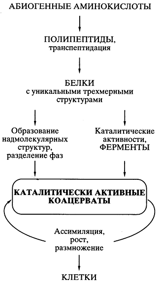
Mal. 5. Schematic representation of the paths of the journey of life
according to the protein-coacervate theory of A.I. Oparina
Everything was well thought out and scientifically grounded in theory, except for one problem, for a long time the eyes of all the fahivts in the gallery were flattened for a long time. Якщо спонтанно, шляхом випадкових безматричних синтезів у коацерваті виникали поодинокі вдалі конструкції білкових молекул (наприклад, ефективні каталізатори, що забезпечують перевагу даному коацервату в зростанні та розмноженні), то як вони могли копіюватися для поширення всередині коацервату, а тим більше для передачі коацерватам- Теорія appeared untimely to propagate the problem of the exact creation - in the middle of the coacervate and in the generations - of single effective protein structures, which appeared out of the blue.
Light RNA as a front daily life. Accumulated knowledge about genetic code, nucleic acids and the biosynthesis of proteins led to the confirmation of a fundamentally new idea about TOM, that everything started not from proteins, but from RNA [-]. Нуклеїнові кислоти є єдиним типом біологічних полімерів, макромолекулярна структура яких завдяки принципу комплементарності при синтезі нових ланцюгів (докладніше див.) забезпечує можливість копіювання власної лінійної послідовності мономерних ланок, іншими словами, можливість відтворення (реплікації) полімеру, його мікроструктури. Therefore, only nucleic acids, but not proteins, can be a genetic material, that is, molecules that repeat their specific microstructure in generations.
It is the low level of RNA itself, and not DNA, that could be the first genetic material.
First, i in chemical synthesis, i in biochemical reactions, ribonucleotides are converted to deoxyribonucleotides; Deoxyribonucleotides are products of modification of ribonucleotides (div. Fig. 2).
In a different way in the most recent, universal processes of life metabolism, ribonucleotides, rather than deoxyribonucleotides, are widely represented, including the main energy sources of the ribonucleoside-polyphosphate (ATP lean) type.
Thirdly, RNA replication can be carried out without any involvement of DNA, and the mechanism of DNA reduplication can be found in the living world in order to influence the overall role of the RNA primer in the initiation of DNA lance synthesis.
Fourthly possessing all the same matrix and genetic functions, such as DNA, RNA, are also capable of low functions, powerful proteins, including catalysis of chemical reactions. In this manner, it is possible to consider DNA as a novel evolutionary approach - as a modification of RNA, specialized for the purpose of victorious functions and the creation of unique copies of genes in the warehouse of the clitin genome without the uninterrupted participation of biosynthesis.
In addition, as soon as catalytically active RNA was used, the idea of the primacy of RNA in life took a strong lead to development, and the concept was formulated self-sufficient world of RNA, scho blowing the current life [ , ]. A possible scheme of RNA viknennia is shown in fig. 6.
The abiogenic synthesis of ribonucleotides in the same covalent association in the oligomer and polymer of the RNA type could be observed approximately in the same minds and in the same chemical conditions that were postulated for the synthesis of amino acids and polypeptides. Recently O.B. Chetverin and his colleagues (Institute of Protein RAS) experimentally showed that polyribonucleotides (RNA) are used in the primary water environment before spontaneous recombination, so that the exchange of lanceolates by the path of transesterification. The exchange of short lancet ribs for a long time is responsible for the reduction of polyribonucleotides (RNA), and the recombination itself is similar to the structural diversity of these molecules. Among them, catalytically active RNA molecules could be blamed.
To bring about the appearance of single RNA molecules, which could catalyze the polymerization of ribonucleotides or the splicing of oligonucleotides on the complementary lance as on the matrix [ ], meant the formation of the mechanism of RNA replication. Replication of the RNA catalysts (ribozymes) themselves is of little consequence in causing self-replicating RNA populations. Producing their own copies of RNA multiplied. Inevitable pardons in copying (mutation) and recombination in RNA populations, which self-replicate, created more and more diversity in the whole world. In this rank, the transfer of the ancient world of RNA - ce "a self-sufficient biological world, in which RNA molecules functioned as genetic material, and as enzyme-like catalysts" .
Vindication to protein biosynthesis. Далі на основі світу РНК мало відбуватися становлення механізмів біосинтезу білка, поява різноманітних білків із успадкованою структурою та властивостями, компартменталізація систем біосинтезу білка та білкових наборів, можливо, у формі коацерватів та еволюція останніх у клітинні структури – живі клітини (див. рис. 6) . ).
The problem of the transition from the ancient world of RNA to the current protein-synthesizing world is important for a fundamentally theoretical solution. Possibility of abiogenic synthesis of polypeptides and protein-like speeches does not help to solve the problem, there are no signs of any specific path, as the synthesis of mig bibuty coats from RNA and under genetic control. Genetically controlling the synthesis of polypeptides and proteins develops independently from the primary abiogenic synthesis, in its own way, with the improvement of already existing RNA. In the literature, a few hypotheses have been proposed for the modern mechanism of protein biosynthesis in the world of RNA, but, perhaps, they can be seen as thought out in detail and without a glance of physical and chemical capabilities. I submit my version of the process of evolution and specialization of RNA, which led to the end of the protein biosynthesis apparatus (small 7), but I do not pretend to be finished.
A hypothetical scheme has been proposed to avenge two hundred moments, which seem to be important.
First, It is postulated that the abiogenically synthesized oligoribonucleotides were actively recombined behind the additional mechanism of spontaneous non-enzymatic transesterification, leading to the reduction of RNA lancets and giving rise to their diversity. In the same way, in the population of oligonucleotides and polynucleotides, both catalytically active RNA species (ribozymes) and other RNA species with specialized functions could appear (Fig. 7). Moreover, non-enzymatic recombination of oligonucleotides that bind complementary to the polynucleotide template could ensure the fusion (splicing) of fragments complementary to this template into a single lance. In this way, instead of catalyzing the polymerization of mononucleotides, the primary copying (multiplication) of RNA could be effected. Assuming that they were ribozymes that had little polymerase activity, then the efficiency (accuracy, flexibility and productivity) of copying on complementary. matrices are small and grow significantly.

Mal. 7. Scheme of evolution and specialization of RNA molecules
in the process of transition from the ancient world of RNA to to the current world
genetically determined protein biosynthesis
Other An important point in my version is that the first apparatus for the biosynthesis of protein appeared in the eradication of many species of specialized RNA before the appearance of the apparatus for enzymatic (polymerase) replication of genetic material - RNA and DNA. Their primary apparatus, including catalytically active proribosomal RNA, has low peptidyl transferase activity; recruitment of pro-tRNAs that specifically bind amino acids or short peptides; other proribosomal RNA, interacting simultaneously with catalytic proribosomal RNA, pro-mRNA and pro-tRNA (div. Fig. 7). Such a system could already synthesize lanceuge polypeptides via a catalyzed transpeptidation reaction. Among the other catalytically active proteins - the primary enzymes (enzymes) - there were also proteins that catalyze the polymerization of nucleotides - replicas, or NK polymerases.
Vtim, perhaps, what is the hypothesis about ancient light RNA, as a leader of today's living world, cannot be sufficient enough to substantiate the main problem - a scientifically plausible description of the mechanism of transition from RNA and replication to protein biosynthesis. The alternative hypothesis of A.D. Альтштейна (Інститут біології гена РАН), в якій постулюється, що реплікація генетичного матеріалу та його трансляція - синтез білка - виникали і еволюціонували одночасно і поєднано, починаючи з взаємодії абіогенно синтезованих олігонуклеотидів і аміноацил-нуклеотидилатів - змішаних ангідридів. Alas, the Cossack is already coming ... ( "I Shahrazad caught the wound, and she pinned the permission to promo".)
Literature
1. Watson JD, Crick F.H.C. Molecular structure of nucleic acids // Nature. 1953. V. 171. P. 738-740. 2. Watson JD, Crick F.H.C. Genetic implications of the structure of deoxyribose nucleic acids // Nature 1953 V. 171. P. 964-967. 3. Spirin A.S. Modern biology and biological safety // Bulletin of the Russian Academy of Sciences. 1997. No. 7. 4. Spirin A.S. On the macromolecular structure of natural high-polymer ribonuclear chemistry // Journal of Molecular Biology. 1960. V. 2. P. 436-446. 5. Kirn S.H., Suddath F.L., Quigley GJ. that in. Three-dimensional tertiary structure of yeast phenylalanine transfer RNA // Science. 1974. V. 185. P. 435-40. 6. Robertas J.D., Ladner J.E., Finch J.T. that in. Structure of yeast phenylalanine tRNA at 3 A resolution // Nature. 1974. V. 250. P. 546-551. 7. Vasiliev V.D., Serdyuk I.N., Gudkov A.T., SPIRin A.S. Self-organization of ribosomal RNA // Sturcture, Function and Genetics of Ribosomes / Eds. Hardesty B. and Kramer G. New York: Springer-Verlag, 1986, pp. 129-142. 8. Baserga SJ., Steitz J.A. Raznomanіtny svіt malikh ribo-nucleoproteins // The RNA World / Eds. Gesteland R.F. and Atkins J.F. New York: Cold Spring Harbor Laboratory Press, Cold Spring Harbor, 1993, pp. 359-381. 9. Kruger K., Grabowski PJ., Zaug AJ. that in. Self-splicing RNA: Autoexcision and autocyclization of the ribosomal RNA intervening sequence of Tetrahymena// cell. 1982. V. 31. P. 147-157. 10. Guerrier-Takada C., Gardiner K., Marsh T. et al. RNA moiety ribonucleases P is a catalytic subunit of enzyme // Cell. 1983. V. 35. P. 849-857. 11. Oparin A.I. Walking life. M: Moscow Robotnik, 1924. 12. Oparin A.I. Vinnyknennya life of the Earth (3rd kind.). M: View of the Academy of Sciences of the SRSR, 1957. 13. Woese S. Evolution of the genetic code // The Genetic Code. New York: Harper & Row, 1967, pp. 179-195. 14. Crick F.H.C. origin of the genetic code // Journal of Molecular Biology. 1968. V. 38. P. 367-379. 15. Orgel L.E. Evolution of the genetic apparatus // Journal of Molecular Biology. 1968. V. 38. P. 381-393. 16. Gilbert W. The RNA world // Nature. 1986. V 319 P. 618. 17. Joyce G.F., Orgel L.E. Prospects for understanding the origin of RNA world // The RNA World / Eds. Gesteland R.F. and Atkins J.F. New York: Cold Spring Harbor Laboratory Press, Cold Spring Harbor, 1993 P 1-25. 18. Chetverina H.V., Demidenko A.A., Ugarov V.I., Chetverin A.B. Spontaneous reorganization in RNA sequences // FEBS Letters. 1999. V. 450. P. 89-94. 19. Bartel D.P., Szostak J.W. Isolation of new ribozymes from a large pool of random sequences // Science. 1993. V. 261. P. 1411-1418. 20. Ekland E.H., Bartel D.P. RNA-catalysed RNA polymerization with nucleoside triphosphates // Nature. 1996 V. 382. P. 373-376. 21. Orgel L.E. origin of life - Review of facts and speculations // Trends in Biochemical Sciences. 1998. V. 23. p. 491-495. 22. Altstein A.D. Pohodzhennya genetic system: the progeny hypothesis // Molecular Biology. 1987. T. 21. S. 309-322.